Welcome to the Amira-Avizo Software Use Case Gallery
Below you will find a collection of use cases of our 3D data visualization and analysis software. These use cases include scientific publications, articles, papers, posters, presentations or even videos that show how Amira-Avizo Software is used to address various scientific and industrial research topics.
Use the Domain selector to filter by main application area, and use the Search box to enter keywords related to specific topics you are interested in.
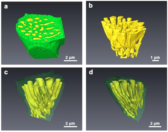
A combination of 3D tomography techniques provides direct evidence that links the phase assemblage to the bio-corrosion behaviour of a magnesium-zinc-calcium (Mg-Zn-Ca) alloy. The true shape, connectivity and volume fraction of the components have been determined using focused ion beam (FIB) tomography. The mechanism of the effect of the components with different morphologies on biodegradation was studied with the help of FIB. Micro X-ray CT (MicroCT) was used to investigate the degradation o... Read more
Y.Lu, R.G.Ding, Y.L.Chiu, I.P.Jones
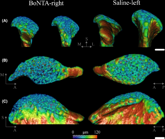
Masseter muscle function influences mandibular bone homeostasis. As previously reported, bone resorption markers increased in the mouse mandibular condyle two days after masseter paralysis induced with botulinum toxin type A (BoNTA), followed by local bone loss.
This study aimed to evaluate the bone quality of both the mandibular condyle and alveolar process in the mandible of adult mice during the early stage of a BoNTA‐induced masseter muscle atrophy, using a combined 3D histomorpho... Read more
Julián Balanta‐Melo, María Angélica Torres‐Quintana, Maximilian Bemmann, Carolina Vega, Constanza González, Kornelius Kupczik, Viviana Toro‐Ibacache, Sonja Buvinic
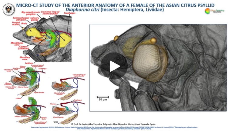
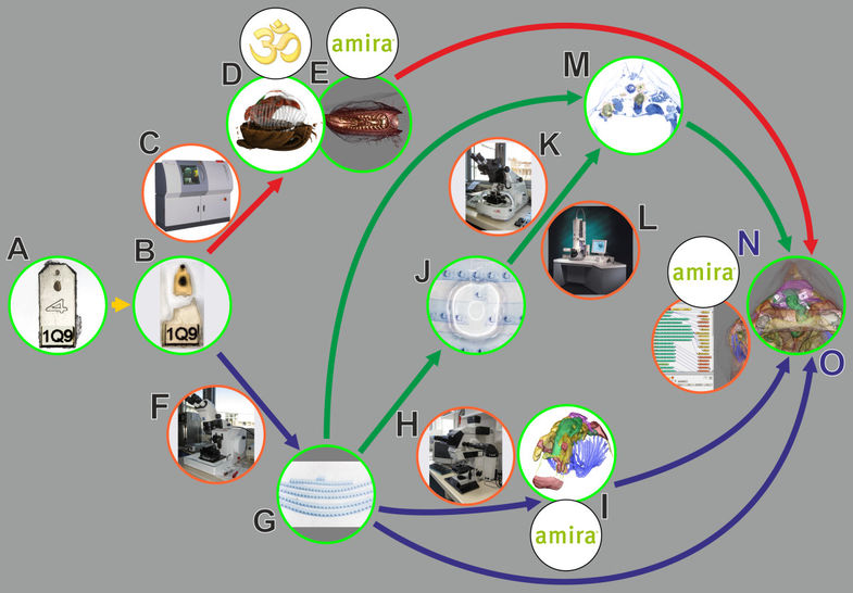
The Asian citrus psyllid (ACP), Diaphorina citri, is a harmful pest of citrus trees that transmits Candidatus Liberibacter spp. which causes Huanglongbing (HLB) (citrus greening disease); this is considered to be the most serious bacterial disease of citrus plants.
Here we detail an anatomical study of the external and internal anatomy (excluding the reproductive system) using micro-computed tomography (micro-CT). This is the first complete 3D micro-CT reconstruction o... Read more
Javier Alba-Tercedor, Wayne B. Hunter & Ignacio Alba-Alejandre
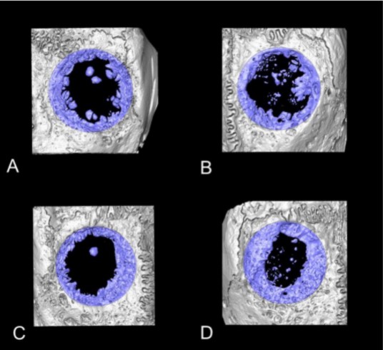
Collagen-Based Matrices for Osteoconduction: A Preclinical In Vivo Study
The aim of this study was to evaluate the influence of additional hydroxyapatite (HA) in collagen-based matrices (CM) and membrane placement on bone formation in calvarial defects.
Critical size defects in the calvaria of 16 New Zealand White Rabbits were randomly treated with CM or mineralized collagen-based matrices (mCM). Half of the sites were covered with a collagen membrane. Animals were euthanized after 12 weeks of healing. The samples were studied by micro-CT and histology. New... Read more
Hiroki Katagiri, Yacine El Tawil, Niklaus P. Lang, Jean-Claude Imber, Anton Sculean, Masako Fujioka-Kobayashi and Nikola Saulacic
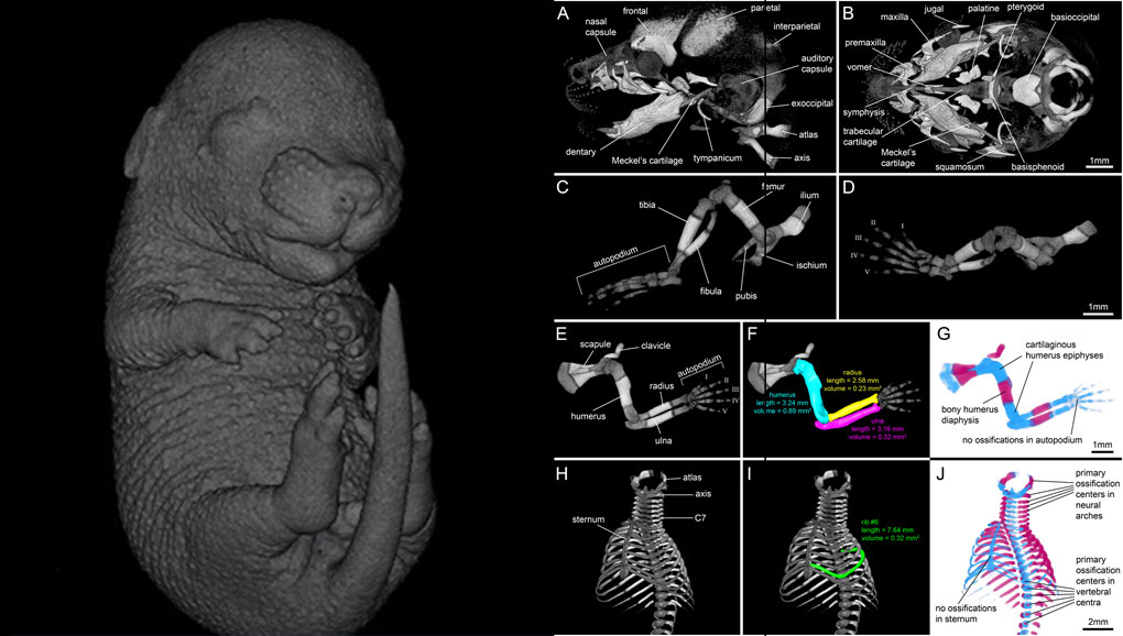
For decades, clearing and staining with Alcian Blue and Alizarin Red has been the gold standard to image vertebrate skeletal development. Here, we present an alternate approach to visualise bone and cartilage based on X-ray microCT imaging, which allows the collection of genuine 3D data of the entire developing skeleton at micron resolution.
Our novel protocol is based on ethanol fixation and staining with Ruthenium Red, and efficiently contrasts cartilage matrix, as demonstrated in wh... Read more
Simone Gabner, Peter Böck, Dieter Fink, Martin Glösmann, Stephan Handschuh
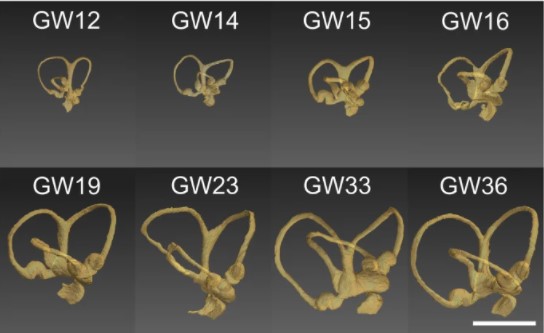
Progressive transformation of the otic placode into the functional inner ear during gestational development in humans leads to the acquisition of hearing perception via the cochlea and balance and spatial orientation via the vestibular organ.
Using a correlative approach involving micro-computerized tomography (micro-CT), transmission electron microscopy and histological techniques we were able to examine both the morphological and cellular changes associated with human inner ear devel... Read more
Lejo Johnson Chacko, David Wertjanz, Consolato Sergi, Jozsef Dudas, Natalie Fischer, Theresa Eberharter, Romed Hoermann, Rudolf Glueckert, Helga Fritsch, Helge Rask-Andersen, Anneliese Schrott-Fischer & Stephan Handschuh
In biomedical research, a huge variety of different techniques is currently available for the structural examination of small specimens, including conventional light microscopy (LM), transmission electron microscopy (TEM), confocal laser scanning microscopy (CLSM), microscopic X-ray computed tomography (microCT), and many others. Since every imaging method is physically limited by certain parameters, a correlative use of complementary methods often yields a significant broader range of inform... Read more
Stephan Handschuh, Natalie Baeumler, Thomas Schwaha & Bernhard Ruthensteiner
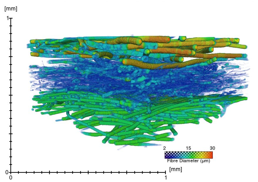
A virtual testing platform for filter materials and textile masks
In order to provide the population with consistent and science-based advice on preferred materials for face masks, we are characterizing the microstructure of different materials using X-ray microfocus computed tomography (microCT), and we use these datasets to simulate the pressure drop (i.e. measure for breathability). We validate our measurements with physically measured filter efficiency and pressure drop, and in this way, we try to develop a “virtual testing platform” for the charac... Read more
The ContrasTTeam of Prof. dr. Greet Kerckhofs, UCLouvain and MTM
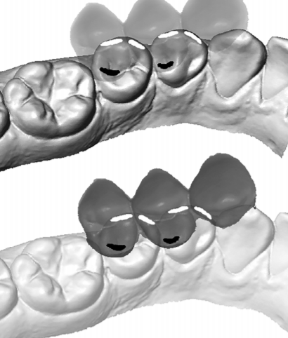
Variation of 3D outer and inner crown morphology in modern human mandibular premolars
This study explores the outer and inner crown of lower third and fourth premolars (P3, P4) by analyzing the morphological variation among diverse modern human groups.
We studied three‐dimensional models of the outer enamel surface and the enamel–dentine junction (EDJ) from μCT datasets of 77 recent humans using both an assessment of seven nonmetric traits and a standard geometric morphometric (GM) analysis. For the latter, the dental crown was represented by ... Read more
Viktoria A. Krenn, Cinzia Fornai, Lisa Wurm, Fred L. Bookstein, Martin Haeusler, Gerhard W. Weber
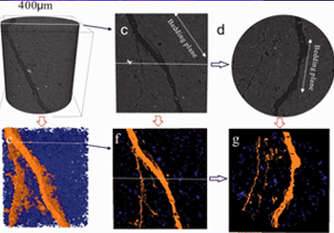
Experimental study on the cracking process of layered shale using X-ray microCT
The cracking process in Longmaxi formation shale was experimentally studied during uniaxial compressive loading. Both the evolution of the three-dimensional fracture network and the micromechanics of failure in the layered shale were examined as a function of the inclination angle of the bedding plane. To visualize the cracking process, the test devices presented here used an industrial X-ray CT scanner that enabled scanning during the uniaxial compressive loading. Scanning electron microscop... Read more
Institue of Geomechanic, Chinese Academy of Geological Sciences, Laboratory of Shale Oil & Gas, Beijing, China
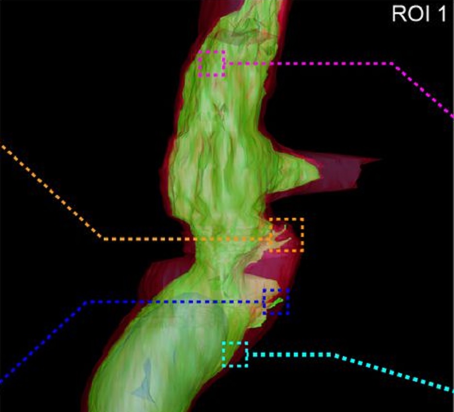
Fast and precise targeting of single tumor cells in vivo by multimodal correlative microscopy
Intravital microscopy provides dynamic understanding of multiple cell biological processes, but its limited resolution has so far precluded structural analysis. Because it is difficult to capture rare and transient events, only a few attempts have been made to observe specific developmental and pathological processes in animal models using electron microscopy. The multimodal correlative approach that we propose here combines intravital microscopy, microscopic X-ray computed tomography and thr... Read more
Matthia A. Karreman, Luc Mercier, Nicole L. Schieber, Gergely Solecki, Guillaume Allio, Frank Winkler, Bernhard Ruthensteiner, Jacky G. Goetz, Yannick Schwab
In biomedical research, a huge variety of different techniques is currently available for the structural examination of small specimens, including conventional light microscopy (LM), transmission electron microscopy (TEM), confocal laser scanning microscopy (CLSM), microscopic X-ray computed tomography (microCT), and many others. Since every imaging method is physically limited by certain parameters, a correlative use of complementary methods often yields a significant broader range of inform... Read more
Stephan Handschuh, Natalie Baeumler, Thomas Schwaha and Bernhard Ruthensteiner
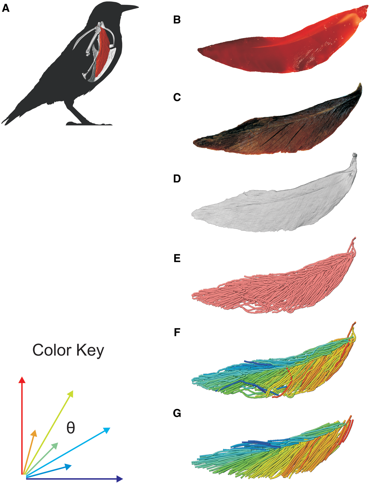
3D Muscle Architecture of the Pectoral Muscles of European Starling (Sturnus vulgaris)
Avian flight is achieved through a number of modifications to the body, including the pectoral girdle (…). Muscle architecture is a critical variable in determining the biomechanical function of the vertebrate musculoskeletal system; however, accurate three-dimensional (3D) understanding of muscle architecture has been historically difficult to acquire. Here, we present a musculoskeletal model of a European starling (Sturnus vulgaris) pectoral girdle generated from iodine contr... Read more
S.P. Sullivan, F.R. McGechie, K.M. Middleton, C.M. Holliday
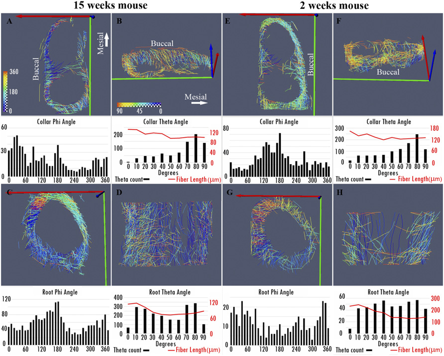
Nonuniformity in ligaments is a structural strategy for optimizing functionality
Ligaments serve as compliant connectors between hard tissues. In that role, they function under various load regimes and directions. The 3D structure of ligaments is considered to form as a uniform entity that changes due to function. The periodontal ligament (PDL) connects the tooth to the bone and sustains different types of loads in various directions. Using the PDL as a model, employing a fabricated motorized setup in a microCT, we demonstrate that the fibrous network structure with... Read more
Gili R. S. Naveh, Jonathan E. Foster, Tomas M. Silva Santisteban, Xianrui Yang, and Bjorn R. Olsen
The Faroe Islands are a group of islands in the North Atlantic that are known for its natural beauty, Viking culture and a special population of house mouse. The Faroese house mouse from the most remote island of the Faroese, Mykines (population: 10 people), looked so distinct when it was discovered that it was declared a subspecies, Mus musculus faeroenesis. These mice are large-bodied and showed an extreme form of left-right asymmetry in its skull. Our research group has... Read more
Yingguang Frank Chan, William H. Beluch, Rémi Blanc
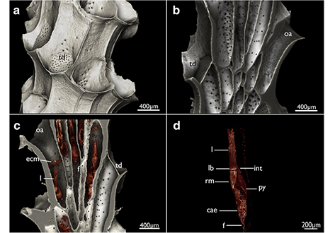
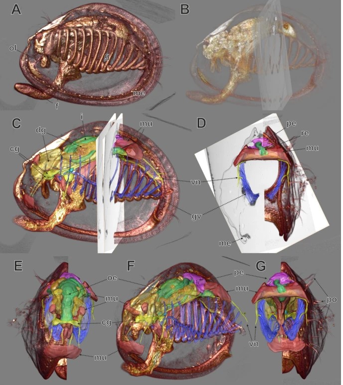
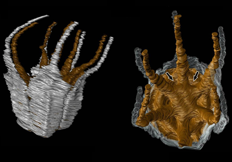
Cyclostome bryozoans are an ancient group of marine colonial suspension-feeders comprising approximately 700 extant species. Previous morphological studies are mainly restricted to skeletal characters whereas data on soft tissues obtained by state-of-the-art methods are still lacking. In order to contribute to issues related to cyclostome ground pattern reconstruction, we analyzed the morphology of the neuromuscular system Cinctipora elegans by means of immunocytochemical staining,... Read more
Thomas F. Schwaha, Stephan Handschuh, Andrew N. Ostrovsky, Andreas Wanninger
Angiosperm-dominated floras of the Late Cretaceous are essential for understanding the evolutionary, ecological, and geographic radiation of flowering plants.
The Late Cretaceous–early Paleogene Deccan Intertrappean Beds of India contain angiosperm-dominated plant fossil assemblages known from multiple localities in central India. Numerous monocots have been documented from these assemblages, providing a window into an important but poorly understood time in their diversification. On... Read more
Kelly K.S. Matsunaga, Selena Y. Smith, Steven R. Manchester, Dashrath Kapgate, Deepak Ramteke, Amin Garbout, and Herminso Villarraga-Gómez
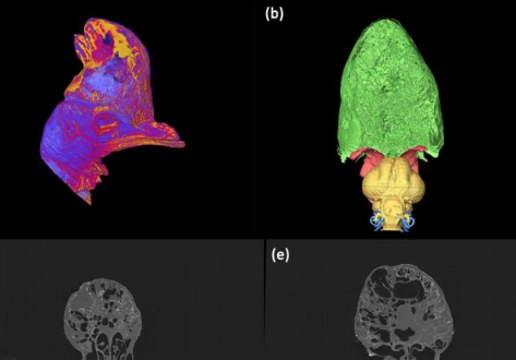
Cranial ornaments such as keratinous horns and bony casques are commonplace amongst birds and take a variety of diverse forms. Possible functions include display, thermoregulation, vocalisation and intraspecific combat, yet few hypotheses have been directly tested. Here we investigate the anatomy and mechanics of the casque of the Southern Cassowary (Casuarius casuarius), and test functional hypotheses using CT-based virtual dissection.
In particular, we determine the nature of pneumat... Read more
Charlotte A. Brassey , Thomas O’Mahoney
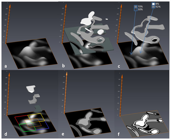
MIA-Clustering: a novel method for segmentation of paleontological material
Paleontological research increasingly uses high-resolution micro-computed tomography (μCT) to study the inner architecture of modern and fossil bone material to answer important questions regarding vertebrate evolution. This non-destructive method allows for the measurement of otherwise inaccessible morphology. Digital measurement is predicated on the accurate segmentation of modern or fossilized bone from other structures imaged in μCT scans, as errors in segmentation can result in inaccur... Read more
Christopher J. Dunmore, Gert Wollny, Matthew M. Skinner